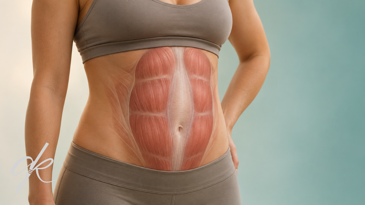
Diastasis recti is a separation of the abdominal muscles at the midline — but it is not a hernia, because there is no defect in the fascia. There is no settled answer to “how many centimetres is normal”: in a study of 1,840 women the mean separation at the standard measurement site was 2.37 cm, meaning the widely cited 2 cm cutoff sits below the population mean. And the bulge does not come from the distance itself but from thinning of the linea alba.
Months after giving birth, the bulge that remains in the mirror is often described in one sentence:
“I’ve lost the weight, but my stomach still looks pregnant.”
Diastasis recti is often behind that picture. But a significant part of what circulates about it — particularly the centimetre thresholds and the back pain link — is not supported in the literature.
This article addresses four questions with their sources: what it is, how it is measured, what exercise actually does, and where surgery comes into the discussion.
What diastasis recti is — and what it is not
The definition
In the front abdominal wall, the two rectus abdominis muscles meet at the midline through a band of connective tissue called the linea alba. Sources note that the rectus muscles are normally separated at the midline by no more than 1 to 2 cm.
Diastasis recti is defined as an abnormal separation of these two muscles resulting from thinning and widening of the linea alba.
🔴 And the crucial distinction: it is not a hernia
Diastasis recti is an acquired condition in which the rectus muscles are separated by an abnormal distance along their length — but with no fascial defect.
That is the fundamental difference from a hernia. In a hernia there is a genuine opening in the abdominal wall through which tissue can protrude. In diastasis there is no opening; the connective tissue has thinned and widened.
| Diastasis recti | Hernia | |
|---|---|---|
| Fascial defect | Absent | Present |
| Mechanism | Thinning and widening of the linea alba | A true defect in the wall |
| Appearance | Diffuse bulge along the midline | Usually a localised protrusion |
This is not a cosmetic detail — as below, whether the two occur together changes the treatment approach.
The bulge does not come from the distance
This section answers the question most people actually have, and it is missing from most accounts.
The bulge associated with diastasis is due to attenuation of the linea alba between the paired muscles, and may occur irrespective of the distance.
What follows from this
Two people with the same measured distance can look different, because what determines appearance is not the number of centimetres between the muscles but how thin and how lax the connective tissue between them has become.
A thin, lax linea alba bulges forward under intra-abdominal pressure. That bulging can be pronounced even when the distance is smaller.
So a measurement — on its own — does not explain the severity of what you see.
How is it measured, and how many centimetres is “normal”?
Method and sites
Measurement is taken as the distance between the medial borders of the two rectus muscles — the inter-recti distance (IRD). Three sites are established in the literature:
- At the xiphoid (the lower tip of the breastbone)
- 3 cm above the umbilicus
- 2 cm below the umbilicus
Ultrasound can be used; some studies have used palpation and a tape measure.
Note: The widely known “finger-width” method gives a rough impression but is not a measurement standard. One study cited two finger-widths as roughly 25 mm — but since finger thickness varies between people, the method does not produce a figure.
Threshold values across sources
| Source / approach | Threshold given |
|---|---|
| Common convention | Up to 2 cm physiological at 3 cm above the umbilicus |
| European Hernia Society guideline | 2 cm or less might be considered physiologically normal |
| Site-specific criteria | >15 mm at the xiphoid, >22 mm at 3 cm above the umbilicus, >16 mm at 2 cm below |
| A cross-sectional study | May be considered normal up to 34 mm at 3 cm above the umbilicus |
| 1948 anatomical data | Linea alba width up to 9-14 mm cranially, up to 18 mm at the umbilicus |
| An ultrasound study | At 2 cm above the navel: 15 mm in parous women, 10 mm in nulliparous |
A reference source summarises the position: there is controversy regarding what constitutes a typical inter-recti distance, at what level measurements should be taken and by what means — and thus when the distance can be considered abnormal.
🔴 And the population data changes the picture
A population study in which 1,840 women were assessed by ultrasound addresses the threshold question directly. Mean age was 35; women less than six months postpartum, exclusively breastfeeding, or with previous midline abdominal surgery, hernia repair or abdominoplasty were excluded.
| Measure | Result |
|---|---|
| Mean IRD at 3 cm above the umbilicus | 2.37 cm (SD 1.19; range 0.35-9.86) |
| 80th percentile at that site | 3.24 cm |
| Mean maximal IRD from any site | 2.77 cm (SD 1.30) |
| Median | 2.59 cm |
| Interquartile range | 1.85-3.47 cm |
| Range | 0.39-12.63 cm |
The authors’ conclusion
Fixed IRD cutoffs commonly used to define diastasis recti fall within the population distribution, with values up to approximately 5 cm corresponding to the 95th percentile.
What that means: the frequently used 2 cm cutoff sits below the population mean at the standard measurement site (2.37 cm).
Applied as a boundary, that threshold classifies a substantial share of women with no complaints at all as “abnormal”.
⚠️ But this does not make measurement pointless. Measurement is meaningful for monitoring and for surgical planning. What it says is different: a figure in centimetres does not on its own make a diagnosis or decide a treatment.
⚠️ The limits of this finding
- The age range was narrow (33-37), so it cannot be generalised to all age groups.
- The first six months postpartum were excluded — so it does not represent the early postpartum period.
- It is a cohort from a single country.
- I could not reach the full text; the figures come from the study’s abstract.
Can exercise fix it?
This is the most contested topic in the literature. Rather than giving a clean answer, I report the sources as they stand — because they contradict each other.
Reviews that found an effect
A meta-analysis of nine randomised controlled trials and 450 participants found a significant reduction in the exercise group:
| Comparison | Mean difference |
|---|---|
| Exercise vs control | -8.05 mm (95% CI -10.43, -5.68) |
| Started before 3 months postpartum | -10.2 mm (95% CI -14.94, -5.46) |
| Structured programme vs standard exercise | -9.59 mm (95% CI -13.45, -5.72) |
The second row is notable: reduction was greater in programmes started early.
Another review reported that abdominal muscle training produced a greater reduction in distance measured at rest and during a head lift than no exercise — but the certainty of evidence was graded “low”.
🔴 Reviews that found no effect, or no meaningful one
A systematic review of 16 randomised controlled trials and 698 participants concluded:
- Moderate certainty evidence from six trials (n=161): abdominal exercise produced a small reduction — -0.43 cm (95% CI -0.82, -0.05)
- Only 3 of the 16 trials had a low risk of bias
“Conservative interventions do not lead to clinically significant reductions in inter-recti distance in women postnatally, but abdominal exercises may have other physical and psychosocial benefits in the management of DRAM.”
A more recent updated review using trial sequential analysis concluded:
“No evidence to support the effectiveness of exercise-based interventions as a standalone treatment for reducing IRD in postpartum women.”
An earlier review by the same group had graded the evidence as “very low quality”.
How should the contradiction be read?
| Question | Answer |
|---|---|
| Is there a measurable reduction? | In several meta-analyses yes — around 4-8 mm |
| Is that reduction clinically meaningful? | Some reviews say no |
| What is the evidence quality? | Low to moderate; most trials carry a high risk of bias |
| Is it sufficient as a standalone treatment? | The updated review found no supporting evidence |
So the debate is not “does exercise work”; it is “is the measured reduction enough to change appearance and function”.
🔴 But this sentence should not be dropped
Even the review that found no meaningful effect states that abdominal exercises may have other physical and psychosocial benefits.
And a scoping review notes that while exercise is the standard approach to diastasis rehabilitation, there is no consensus on the most effective routine.
So the right framing is not “exercise is pointless”. Exercise may not close the gap; but its value for abdominal wall function, strength and quality of life is a separate assessment.
The European Hernia Society guideline also notes that physiotherapy may be considered before surgical management.
The back pain link: not established
What the systematic review found
| Finding | Proportion |
|---|---|
| Studies finding no association | 61.5% |
| Studies finding a positive correlation | 38.5% |
🔴 And the crucial distinction
Women with diastasis recti tend to have weaker abdominal muscles and a higher prevalence of abdominal pain; but no higher prevalence of pelvic floor disorders, low back pain or pelvic girdle pain than women without it.
| Finding | Status |
|---|---|
| Weaker abdominal muscles | More common |
| Abdominal pain | More common |
| Low back pain | Not more common |
| Pelvic girdle pain | Not more common |
| Pelvic floor dysfunction | Not more common |
Other findings
- One study found no difference in reported lumbopelvic pain between women with and without diastasis (p=0.10).
- A cited study reported that mild diastasis was not associated with lumbopelvic pain, and that women with diastasis were not more likely to report it 12 months postpartum.
- Another study found no association between diastasis and lumbopelvic pain or incontinence.
- A pilot study using ultrasound and an algometer found no significant relationship between increased abdominal distance and increased lumbopelvic pain.
⚠️ The finding in the other direction
For balance: one cross-sectional study reported a significant association between diastasis and lumbopelvic pain in postpartum women (p<0.05).
So the literature is not one-sided — but the weight is on the side finding no association.
What this means for you
If you have back pain, attributing it automatically to diastasis and assuming it will resolve by correcting the diastasis is not supported. Treating back pain as requiring its own assessment is the sounder approach.
By contrast, abdominal pain and abdominal muscle weakness do occur more often alongside diastasis — those two complaints deserve separate attention.
The relationship with hernia
They can occur together
Classification work lists concomitant hernias as a separate heading: umbilical, epigastric, port-site and incisional hernias.
So a genuine opening can also be present within a midline of thinned connective tissue.
🔴 And this changes the approach
The European Hernia Society guideline notes a mesh-based repair in patients with concomitant midline hernias, and a different approach in those without.
So “I have diastasis” does not describe a single clinical picture; whether a hernia accompanies it is decisive.
A relationship in the other direction
A cited assessment notes that diastasis raises the risk of hernia recurrence after midline ventral hernia repair, with recurrence rates reported as high as 31.2% when diastasis is not addressed.
Note: This figure is a report from particular patient groups, not an expectation that transfers directly to every patient.
Is there a centimetre threshold for surgery?
The honest answer: no.
There is a classification — but it is not an indication
A working group of the German Hernia Society and the International Endohernia Society developed a classification proposal:
| Dimension | Categories |
|---|---|
| Width | W1 < 3 cm · W2 = 3-5 cm · W3 > 5 cm |
| Length (sector) | Subxiphoidal, epigastric, umbilical, infraumbilical, suprapubic |
| Concomitant hernia | Umbilical, epigastric, port-site, incisional |
| Other | Previous operations, number of pregnancies and multiple births, skin condition, severity and localisation of preoperative pain |
But this is a classification system — its purpose is to make the results of surgical techniques comparable. The working group states such a classification was needed for the assessment of treatment results in these patients.
So being W2 or W3 does not mean “surgery is required”. A classification is not an indication.
🔴 And the guidelines’ own cautions
- “None of the existing classification systems seems to be optimal for classifying RD.”
- Literature on the definition, classification, symptoms, outcomes and treatments was limited in quality, leading to weak recommendations for the majority of the key questions.
- There is limited evidence on an exact definition.
The working group that developed the classification also notes that published literature on surgical treatment is assessed in systematic reviews as being of low scientific and methodological quality, and that based on current literature no clear distinction can be made in recurrence rate, postoperative complications or patient-reported outcomes.
So what does the decision rest on?
The guideline names the most relevant symptoms as:
Impaired body image and core instability.
Note what stands out: neither is measured in centimetres.
And the guideline notes that physiotherapy may be considered before surgical management.
One practical note
The guideline also notes that in many countries there is no financial cover for the surgical repair of diastasis presented as a solitary condition.
Worth discussing at consultation
- What is my actual complaint? Appearance, core instability, or abdominal pain? The guideline names body image and core instability as the most relevant symptoms.
- At which sites and by what method was the measurement taken? Change the site or method and the figure changes.
- Is there a concomitant hernia? This is the single most important finding for the approach.
- What is the condition of the linea alba? It determines the bulge more than the distance does.
- How long since delivery? Greater reduction has been reported in programmes started early.
- What should I expect from physiotherapy? Discussing the measured reduction and how it translates to appearance builds realistic expectations.
- Should my back pain be assessed separately? The literature does not support attributing it to diastasis.
- What is my skin condition? It appears as a separate heading in the classification.
- Am I planning another pregnancy? This can affect timing.
Summary
Diastasis recti is an abnormal separation of the rectus muscles caused by thinning and widening of the linea alba — but not a hernia, because there is no fascial defect. The distinction matters because whether a hernia accompanies it changes the treatment approach.
There is no settled answer to “how many centimetres”. Common convention treats up to 2 cm at 3 cm above the umbilicus as physiological; but in a study of 1,840 women the mean at that site was 2.37 cm, and the authors noted that commonly used fixed cutoffs fall within the population distribution, with values up to approximately 5 cm corresponding to the 95th percentile. Measurement is meaningful for monitoring and planning, but a figure alone does not make a diagnosis.
The bulge does not come from the distance. Sources state it is due to attenuation of the linea alba and may occur irrespective of the distance — which explains why two people with the same measurement can look different.
On exercise the literature conflicts. A meta-analysis of nine trials reported around 8 mm reduction, and about 10 mm when started early. A review of 16 trials found a small reduction of 0.43 cm but stated conservative interventions do not lead to clinically significant reductions; an updated review found no evidence supporting exercise as a standalone treatment. That same review nonetheless notes abdominal exercises may have other physical and psychosocial benefits, and the guideline notes physiotherapy may be considered before surgery.
The back pain link is not established. In a systematic review, 61.5% of studies found no association. The more precise finding: women with diastasis have weaker abdominal muscles and more abdominal pain, but not more low back pain, pelvic girdle pain or pelvic floor dysfunction.
There is no centimetre threshold for surgery. W1, W2 and W3 are a classification developed to compare outcomes, not to set an indication. The European Hernia Society guideline states none of the existing classification systems seems optimal, that literature quality is limited and that most recommendations are weak. The most relevant symptoms are named as impaired body image and core instability — neither measured in centimetres.
Which approach suits your own situation can be determined by assessing the nature of your complaint, the measurement findings, the condition of the linea alba, whether a hernia is present and how long it has been since delivery. That assessment requires examination by a physician. If you would like your own case assessed, you are welcome to arrange a consultation.
Frequently Asked Questions
Is diastasis recti a hernia?
No. By definition there is no fascial defect in diastasis; the linea alba has thinned and widened. In a hernia there is a genuine opening in the abdominal wall. However the two can occur together, and the presence of a concomitant hernia changes the treatment approach.
How many centimetres of separation is normal?
There is no settled threshold. Common convention treats up to 2 cm at 3 cm above the umbilicus as physiological, and the European Hernia Society guideline notes 2 cm or less might be considered physiologically normal. However, in a study of 1,840 women the mean at that site was 2.37 cm, and the authors noted commonly used fixed cutoffs fall within the population distribution.
How is it measured?
The distance between the medial borders of the two rectus muscles is measured. Established sites are the xiphoid, 3 cm above the umbilicus and 2 cm below. Ultrasound can be used. The finger-width method gives a rough impression but is not a measurement standard.
Why won’t the bulge go away?
Sources state that the bulge associated with diastasis is due to attenuation of the linea alba and may occur irrespective of the distance. So the bulge can be pronounced even when the measured value is small.
Can exercise fix it?
The literature conflicts. A meta-analysis of nine randomised trials reported around 8 mm reduction. A review of 16 trials found a small reduction of 0.43 cm but stated conservative interventions do not lead to clinically significant reductions. An updated review found no evidence supporting exercise as a standalone treatment.
So is exercise pointless?
That does not follow. The review that found no meaningful effect itself notes abdominal exercises may have other physical and psychosocial benefits. The European Hernia Society guideline also notes physiotherapy may be considered before surgical management.
When is the better time to start exercising?
A meta-analysis reported greater reduction in interventions started before 3 months postpartum (around 10.2 mm). However the timing and content of a programme require individual assessment.
Does diastasis cause back pain?
The literature does not support this. In a systematic review, 61.5% of studies found no association between diastasis and low back pain. The more detailed finding is that women with diastasis have weaker abdominal muscles and more abdominal pain, but not more low back pain, pelvic girdle pain or pelvic floor dysfunction.
Above how many centimetres is surgery needed?
There is no established centimetre threshold. W1 (<3 cm), W2 (3-5 cm) and W3 (>5 cm) are a classification system developed to make surgical outcomes comparable, not to set an indication. The European Hernia Society guideline states none of the existing classification systems seems to be optimal.
Then what does the surgical decision rest on?
The guideline names impaired body image and core instability as the most relevant symptoms; neither is measured in centimetres. Concomitant hernia, previous operations, pregnancy history and skin condition also appear as separate headings in the classification.
Does diastasis matter if hernia repair is planned?
A cited assessment notes that diastasis raises the risk of hernia recurrence after midline ventral hernia repair, with rates reported as high as 31.2% when diastasis is not addressed. That figure is a report from particular patient groups.
Op. Dr. Ahmet Kaplan — Specialist in Plastic, Reconstructive and Aesthetic Surgery.
This content is for information only, does not constitute medical advice and does not replace examination by a physician. The measurements, rates and guideline statements cited come from published studies and carry the sample and methodological limitations described in the text; individual situations vary with anatomy, obstetric history, tissue characteristics and clinical circumstances.